Pneumothorax Ct - Tension Pneumothorax Time To Change The Old Mantra Litfl / Findings on ct scan suggestive of pneumothorax include:
byAdmin•
0
Pneumothorax Ct - Tension Pneumothorax Time To Change The Old Mantra Litfl / Findings on ct scan suggestive of pneumothorax include:. A collapsed lung requires immediate medical care. Chest radiography is the first investigation performed to assess pneumothorax, because it is simple, inexpensive, rapid, and noninvasive; Women with catamenial pneumothorax have recurrent episodes of pneumothorax that occur within 72 hours before or after the start of menstruation. Where, a is apex to apex lung. Chest ct is the most reliable imaging study for the diagnosis of pneumothorax, but it is not recommended for routine use in pneumothorax.
Recurrent pneumothorax was diagnosed more frequently in the group with a delayed diagnosis (82% vs. Multidetector ct scan is highly effective in measuring the volume of pneumothoraces. Ct is extremely sensitive for pneumothorax, although controversy remains over the proper management of pneumothoraces seen only on ct (see text). Subpleural blebs and bullae are found at the lung apices at thoracoscopy and on ct scanning in up to 90% of cases of psp, 5 6 and are thought to play a role. Ultrasound imaging also may be used to identify a pneumothorax.
Pneumothorax from www.saem.org Don't be shy to ask for a ct scan in this scenario. Ct scan is the most accurate mean to differentiate the two diagnoses double wall sign described in cases with ruptured bulla causing pneumothorax (air outlining both sides of the bulla wall parallel to the chest wall) lesson: (d) axial ct scan shows extension of the pneumothorax into the retroperitoneum and the soft tissues of the thigh. Recurrent pneumothorax was diagnosed more frequently in the group with a delayed diagnosis (82% vs. Pneumothorax is the medical term for a collapsed lung, a condition in which air or gas is trapped in the space surrounding the lungs causing the lungs to collapse. (a) supine chest radiograph shows a thin white line (white arrows) associated with the deep sulcus sign (black arrows), in keeping with basal or subpulmonic pneumothorax. Size is of less importance than clinical severity (hypoxia, distress, pain, hemodynamics) and pneumothorax size should not dictate management on its own. 3 thus, while chest radiography is not as effective as ct scanning, it does still provide a useful and reasonably accurate estimate of pneumothorax size in most cases using the method outlined in the current guidelines.
This imaging modality can help to accomplish the following:.
This imaging modality can help to accomplish the following:. They are sometimes mistaken for pneumothorax. A collapsed lung requires immediate medical care. Collapsed lung (pneumothorax) a collapsed lung occurs when air gets inside the chest cavity (outside the lung) and creates pressure against the lung. Check out pneumothorax on ebay. Formula to calculate percentage of pneumothorax: Size is of less importance than clinical severity (hypoxia, distress, pain, hemodynamics) and pneumothorax size should not dictate management on its own. Ct was performed in all of the patients who were diagnosed promptly, but in none of the patients with a delayed diagnosis. 3 thus, while chest radiography is not as effective as ct scanning, it does still provide a useful and reasonably accurate estimate of pneumothorax size in most cases using the method outlined in the current guidelines. Recurrent pneumothorax was diagnosed more frequently in the group with a delayed diagnosis (82% vs. However, it is much less sensitive than chest computed. Where, a is apex to apex lung. Also known as pneumothorax, collapsed lung is a rare condition that may cause chest pain and make it hard to breathe.
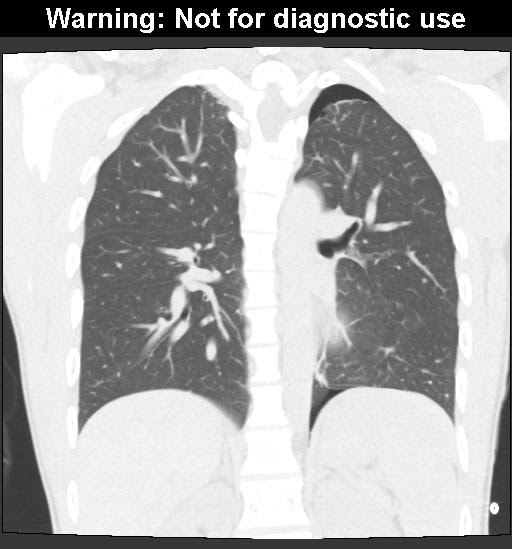
As the free air will tend to collect in the apices, this area of the chest radiograph is examined closely for a possible pneumothorax. Ultrasound imaging also may be used to identify a pneumothorax. We have almost everything on ebay. Ct scan of the chest showing a pneumothorax on the person's left side (right side on the image). Since she remained hemodynamically stable, emergency needle decompression was not necessary.
Pneumothorax And Pneumomediastinum In Covid 19 A Case Series The American Journal Of The Medical Sciences from els-jbs-prod-cdn.jbs.elsevierhealth.com Chest radiography (fig 1) revealed a large left pneumothorax with depression of the left hemidiaphragm, shift of the mediastinum to the right, and left apical blebs. (d) axial ct scan shows extension of the pneumothorax into the retroperitoneum and the soft tissues of the thigh. Chest ct is the most reliable imaging study for the diagnosis of pneumothorax, but it is not recommended for routine use in pneumothorax. Findings on ct scan suggestive of pneumothorax include: (a) supine chest radiograph shows a thin white line (white arrows) associated with the deep sulcus sign (black arrows), in keeping with basal or subpulmonic pneumothorax. Ct is extremely sensitive for pneumothorax, although controversy remains over the proper management of pneumothoraces seen only on ct (see text). Women with catamenial pneumothorax have recurrent episodes of pneumothorax that occur within 72 hours before or after the start of menstruation. Size is of less importance than clinical severity (hypoxia, distress, pain, hemodynamics) and pneumothorax size should not dictate management on its own.
As the free air will tend to collect in the apices, this area of the chest radiograph is examined closely for a possible pneumothorax.
As the free air will tend to collect in the apices, this area of the chest radiograph is examined closely for a possible pneumothorax. Size is of less importance than clinical severity (hypoxia, distress, pain, hemodynamics) and pneumothorax size should not dictate management on its own. The incidence, risk factors, and clinical significance of delayed pneumothorax are not well known. Also known as pneumothorax, collapsed lung is a rare condition that may cause chest pain and make it hard to breathe. But did you check ebay? Chest radiography is the first investigation performed to assess pneumothorax, because it is simple, inexpensive, rapid, and noninvasive; Multidetector ct scan is highly effective in measuring the volume of pneumothoraces. Tension pneumothorax is an unusual but a recognized cause of respiratory and cardiovascular compromise during anaesthesia and surgery. Ultrasound imaging also may be used to identify a pneumothorax. However, it is much less sensitive than chest computed. When bullous disease is present, a loculated pneumothorax may appear similar. Recurrent pneumothorax was diagnosed more frequently in the group with a delayed diagnosis (82% vs. Findings on ct scan suggestive of pneumothorax include:
Subpleural blebs and bullae are found at the lung apices at thoracoscopy and on ct scanning in up to 90% of cases of psp, 5 6 and are thought to play a role. Provided lung windows are examined, a pneumothorax is very easily identified on ct, and should pose essentially no diagnostic difficulty. Check out pneumothorax on ebay. It is better to do right than be right. Where, a is apex to apex lung.
C31 Pneumothorax Ppt Download from slideplayer.com Chest radiography is the first investigation performed to assess pneumothorax, because it is simple, inexpensive, rapid, and noninvasive; Imaging of pneumothorax on ultrasonography is discussed in more detail separately. Formula to calculate percentage of pneumothorax: A collapsed lung requires immediate medical care. But did you check ebay? Ct scan is the most accurate mean to differentiate the two diagnoses double wall sign described in cases with ruptured bulla causing pneumothorax (air outlining both sides of the bulla wall parallel to the chest wall) lesson: Chest ct is the most reliable imaging study for the diagnosis of pneumothorax, but it is not recommended for routine use in pneumothorax. To estimate the size of a pneumothorax, different modalities can be used, such as chest radiography and ultrasound, with ct scans constituting the standard of reference 3 .
Size is of less importance than clinical severity (hypoxia, distress, pain, hemodynamics) and pneumothorax size should not dictate management on its own.
A pneumothorax can be a complete lung collapse or a collapse of only a portion of the lung. Women with catamenial pneumothorax have recurrent episodes of pneumothorax that occur within 72 hours before or after the start of menstruation. Tension pneumothorax is an unusual but a recognized cause of respiratory and cardiovascular compromise during anaesthesia and surgery. Multidetector ct scan is highly effective in measuring the volume of pneumothoraces. Collapsed lung (pneumothorax) a collapsed lung occurs when air gets inside the chest cavity (outside the lung) and creates pressure against the lung. Findings on ct scan suggestive of pneumothorax include: Formula to calculate percentage of pneumothorax: It is better to do right than be right. We have almost everything on ebay. The wall is usually less than 1 mm in thickness and may even be invisible, making detection of the bullae difficult; Provided lung windows are examined, a pneumothorax is very easily identified on ct, and should pose essentially no diagnostic difficulty. (d) axial ct scan shows extension of the pneumothorax into the retroperitoneum and the soft tissues of the thigh. We suggest that the guideline is an.